Healthcare with Confidence
Herniated disc (also called “pinched nerve”, “bulging disc”, “ruptured disc”, “disc protrusion”) is a pathology of the spinal disc, which inner core is leaking through the damaged portion of the outer part of the disc and puts pressure on the nerve spinal roots, as well as spinal canal that leads to a narrowing of the latter (stenosis).
A herniated disc can form in any section of the spine – the lower back, in the thoracic section (upper back) and in the cervical section.
In order to determine the cause of the pain and make the correct diagnosis contact our experts and send us pictures of MRI or CT of the spine.
The main symptoms of a herniated disc is a pain in the back, buttocks radiates to the legs down to the feet.
Causes
Pinched nerve. Here the disk itself is not painful, but bulging, it exerts pressure on a nerve causing pain called “radicular pain”. This pain irradiates to other body parts, for example, from the lower back down the leg or neck down the arm. Leg pain from pinched nerve is called sciatica. Other common causes of a pinched nerve may be spinal stenosis and bone spurs as a result of spinal arthritis.
Soreness of the disc itself. When the patient has focused pain and / or pain in the legs, the source of pain in the disc space.
Any of the above problems may occur in the cervical, thoracic or lumbar spine.
They tend to be most common in the lower back because lower back bears the greatest daily power load duplicator stencils.
Diagnostics
Medical diagnosis of a herniated disc is focused on determining the cause of the patient’s pain or other symptoms.
The main methods of diagnosis:
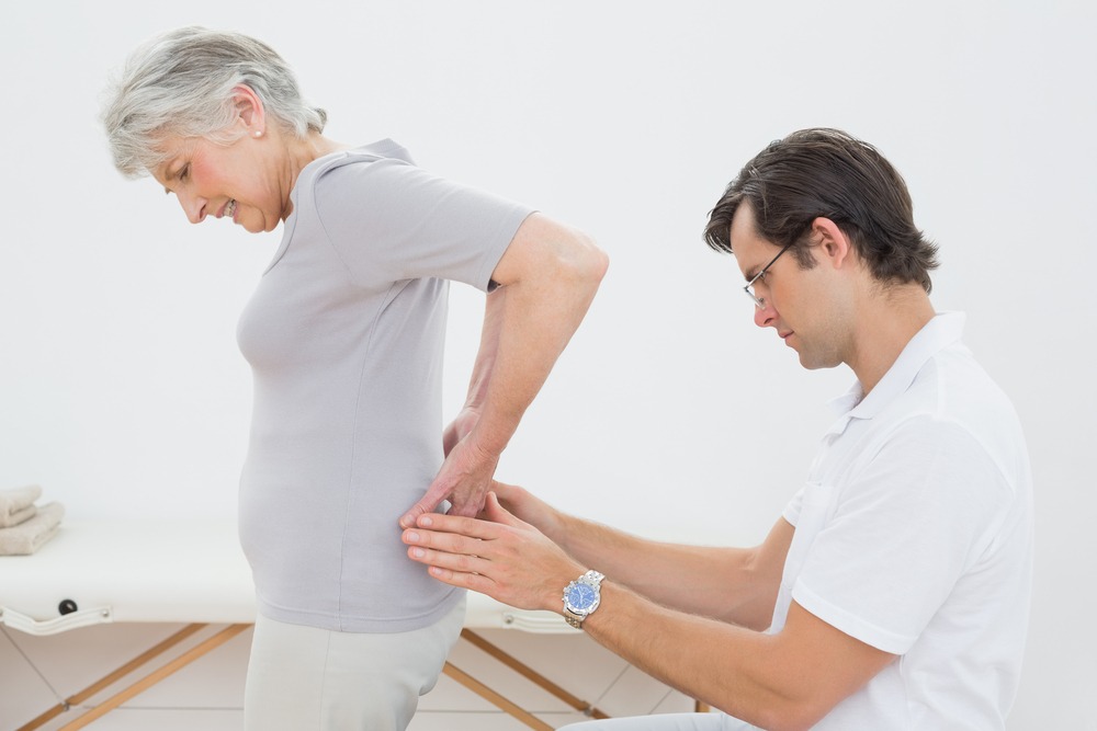
Physical examination. Depending on the symptoms, physical examination may include an assessment of nerve function in certain parts of the feet or hands – tapping a reflex hammer on the different areas. If no response, it may be the full compression of the nerve. Sensory tests can also be carried out using thermal stimulation – hot and cold to determine the response.
Study of muscle strength. In order to determine a deeper degree of pressure on the spine herniation of the spinal nerve, the specialist performs a neurological examination to assess muscle strength, as well as an external examination to detect possible muscle atrophy, muscle twitching, or any abnormal movements.
Pain on palpation or movement of certain areas can also give an idea of what actually produces pain. For example, pain over the sacroiliac joint palpation may indicate a dysfunction of the sacroiliac vertebrae. Pain when straightening the legs may indicate a pinched nerve. Pain with pressure on the lower back may indicate pain from degenerative processes in the disc.
Review of the history of the disease needs to exclude (or determine) other possible causes of pain. The history includes information such as the presence of recurring health problems, previous diagnoses, recent procedures and operations, response to those procedures medication, family history of disease, and any other health problems.
Tests
![]() CT – computed tomography – assessment of the spine by X-rays
CT – computed tomography – assessment of the spine by X-rays
![]() MRI – magnetic resonance imaging – assessment of the spinal nerves and spinal anatomy of the patient, including a disc shape, size, hydration and configuration
MRI – magnetic resonance imaging – assessment of the spinal nerves and spinal anatomy of the patient, including a disc shape, size, hydration and configuration
![]() Discogram – if the cause of the pain is in the disc itself doctor may recommend holding a discogram to determine which disc hurts. During this procedure radiographic contrast material is injected into the disc and cause the reaction of it.
Discogram – if the cause of the pain is in the disc itself doctor may recommend holding a discogram to determine which disc hurts. During this procedure radiographic contrast material is injected into the disc and cause the reaction of it.
Treatment of herniated disc
Only after a thorough examination of the patient experienced professional can be defined treatment plan. For example, the treatment of lumbar disc herniation will not be effective if the actual cause of the patient’s pain is muscle strain or injury to the soft tissues.
Surgery may be considered if the application of non-surgical treatments, such as painkillers, injections, chiropractic and physical therapy is not proven to be effective.
Operation can help in the event that the actual cause of the pain is really a herniated disc or degenerative processes of the disc, which may be seen on MRI.
If one of these conditions is a source of pain the following options for the operation may be considered:
Microdiscectomy – an operation in which part of the disc is removed.
Spinal fusion – surgery for osteochondrosis, which results in the merging of disc space and in which the vertebrae are fixed with special flexible clamps, resulting in the flexibility of the spine is maintained.
The operation is generally not suitable if the cause of the pain is not completely established.
Leading Israeli Spine Surgeons – Second Opinion
Our Doctors


