Healthcare with Confidence
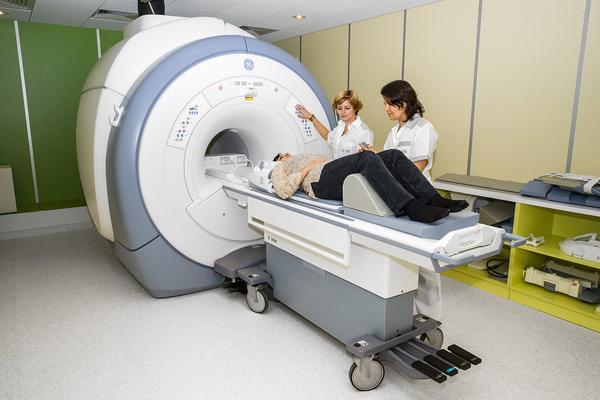
Nuclear magnetic resonance (NMR) or magnetic resonance imaging (MRI) is a medical diagnostic imaging technique used by Israeli doctors for medical diagnosis of various conditions. MRI has no ionizing effect on the body (does not irradiate). MRI scanners use magnetic fields and radio waves to form images of the body.
Israel medicine is designed in such a way that MRI test can be prescribed by an Israeli doctor and performed in one of the largest hospitals in Israel, which has all the necessary professional medical equipment. In Israel you can also get a second opinion MRI / PET / CT from an Israeli doctor radiologist that can be used for confirming / verifying your diagnosis.
Contraindications to MRI include mainly cochlear implants and pacemakers, osteometallic structures and metallic foreign bodies in the body.
MRI of the brain and spinal cord
MRI of the brain and spinal cord is used by Israeli doctors to visualize white matter tracts and malignant diseases of the nervous system. This diagnostics is more sensitive for small tumors than CT and offers better visualization of the posterior fossa. MRI is the optimal choice for diagnosing many central nervous system problems, including demyelinating diseases, dementia, cerebrovascular diseases, infectious diseases, and epilepsy.
Because MRI scans are taken in milliseconds one after another, it allows professionals to see how the brain responds to various stimuli, so that they can study the functional and structural abnormalities of the brain in mental disorders. MRI is also used in navigation in stereotactic surgery and radiosurgery for the treatment of intracranial tumors, arteriovenous malformations, etc.
MRI of the musculoskeletal system
Magnetic nuclear analysis of the musculoskeletal system includes images of the spine and spinal cord, assessment of joint diseases and soft tissue tumors.
MRI of the cardiovascular system (MRA)
MR angiography for congenital heart defects is an adjunct to other imaging techniques such as echocardiography, scintigraphy, and cardiac CT. The addition of angiography includes assessment of myocardial ischemia, cardiomyopathy, myocarditis, iron overload, vascular disease, and congenital heart disease.
MRI of liver and gastrointestinal tract
Hepatobiliary MRI is used by Israeli doctors to detect and evaluate the extent of damage to the liver, pancreas, and bile ducts. Coordination or diffuse liver abnormalities can be assessed using diffusion-weighted images and dynamic contrast enhancement sequences. Extracellular contrast agents are widely used in MRI of the liver, and hepatobiliary contrast agents also provide the ability to provide images of the bile ducts.
Anatomical images of the bile ducts are obtained using a sequence of heavily T2-weighted images in magnetic resonance cholangiopancreatography (MRCP). This is the name of a special protocol for MRI of the pancreas. Functional imaging of the pancreas is performed following secretin administration.
MR enterography provides a non-invasive assessment of inflammatory bowel disease and small bowel tumors. MRI colonography is helpful in identifying large polyps in patients at increased risk of developing colorectal cancer.
Functional MRI
Functional MRI (fMRI) is used to assess how different parts of the brain respond to external stimuli or passive activity at rest. Given the level of oxygenation of the blood, MRI measures the hemodynamic response to short nervous activity as a result of a change in the ratio of oxyhemoglobin to deoxyhemoglobin. To construct a 3D parametric brain map, statistical methods are used that indicate those areas of the brain that show significant changes in activity in response to a task. MRI has applications in behavioral and cognitive research, as well as in complex neurosurgery.
MRI in oncology
For cancer diagnostics MRI is used during preoperative examination, in determining the method of surgery for colorectal cancer, prostate cancer, breast cancer, ovarian cancer, brain cancer, or cervical cancer, and also plays an important role in for systemic treatment decisions.
MRI safety and implants
All patients requiring MRI diagnostics undergo a preliminary consultation for safety. All possible implants in the body fall into several categories:
MR-Safe is a device or implant that is completely non-magnetic, non-conductive, and non-RF reactive, eliminating all primary potential threats during an MRI procedure.
MR-Conditional – A device or implant that may contain magnetic, electrically conductive, or RF-reactive components that are safe for operations in the immediate vicinity of an MRI, provided the conditions for safe use are met.
MR-Unsafe are objects that are significantly ferromagnetic and pose a direct threat to people and equipment inside the magnetic chamber. MRI environment can harm patients with MR-unsafe.
Titanium and titanium alloys are MRI safe.
Before MRI scans of patients with magnetic shunt of the brain, our specialists set it up for a safe mode.
MRI EEG
In the course of research, structural MRI or functional MRI can be combined with EEG (electroencephalography), provided that the EEG equipment (electrodes, amplifiers and peripherals) and MR are compatible.
Contrast agent
The administration of a contrast agent (contrast agent or dye) helps to demonstrate various anatomical structures or pathologies. T1-weighted images of magnetization are used to assess the cerebral cortex, to identify adipose tissue characterizing focal liver lesions, and in general to obtain morphological information.
T2-weighted images of magnetization are used to detect edema and inflammation, detect white matter damage, and evaluate zonal anatomy in the prostate and uterus.
MRI to visualize anatomical structures or blood flow does not require contrast agents because the different properties of tissues or blood provide natural contrast. However, for more specific types of imaging, intravenous contrast agents based on gadolinium chelates are most commonly used. In general, these agents are safer than iodinated contrast agents used in X-ray radiography or CT.
Anaphylactic reactions are very rare, occurring in 0.03-0.1% of cases. Of particular interest is the lower level of nephrotoxicity compared to iodinated agents when administered at usual doses, which has made MRI contrast imaging a feasible option for patients with renal insufficiency who cannot undergo contrast-enhanced CT diagnostics.
Although gadolinium agents have been shown to be safe in patients with renal impairment, patients with severe renal impairment requiring dialysis are at risk of a rare but serious condition, nephrogenic systemic fibrosis, which may be associated with the use of certain gadolinium-containing agents. The most common is gadodiamide.
We follow instructions of the leading Israeli doctors, providing prompt coordination of MRI diagnostics in one of the biggest hospitals mentioned by the doctor, transfer to the clinic, oral and written translations of the results.
Computed tomography is usually prescribed by a doctor in one of the Israeli hospitals, such as Assuta Medical Center, Ichilov, Sheba, Beilinson.
MRI results interpretation is done by an experienced Israeli radiologist, nuclear medicine specialist who has many years of experience in MRI interpretation.
You can get a second opinion of a leading Israeli radiologist on the MRI scan done in your country. Please contact us to clarify details.
MRI types performed in Israeli hospitals
MRI of soft tissues
MRI wrist
MRI knee
MRI maxillofacial joint
MRI thorax and lungs
MRI spine
MRI radiosurgery
MRI hip
MRI ankle
MRI elbow
MRI abdomen enterography
MRI shoulder
MRI forearm
MRI brain
MRA angiography brain
MRI for children (anesthesia)
MRI breast
MRI pancreas protocol (MRCP)
MRI hip
MRI orbits
MRI head
MRI abdomen and pelvis
MRI shoulder


